Arthoscopy Medial Gutter Knee

The Lateral Gutter Drive Through Sign An Arthroscopic Indicator Of Acute Femoral Avulsion Of The Popliteus Tendon In Knee Joints Sciencedirect
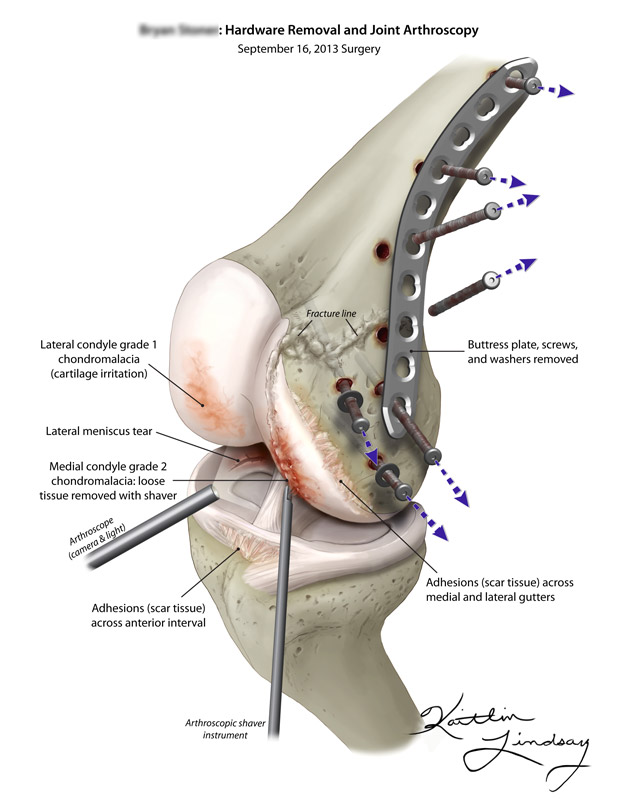
Knee Arthroscopy Hardware Removal Kaitlin Lindsay

Knee Diagnostic Arthroscopy In Left Knee After Gunshot Wound The Download Scientific Diagram

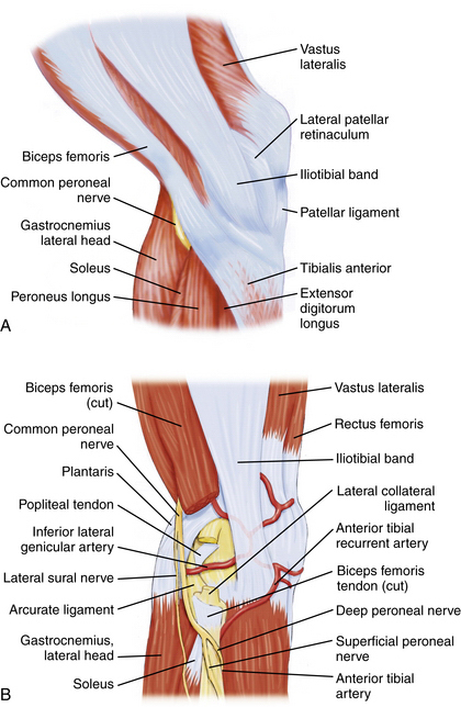
The Knee Musculoskeletal Key

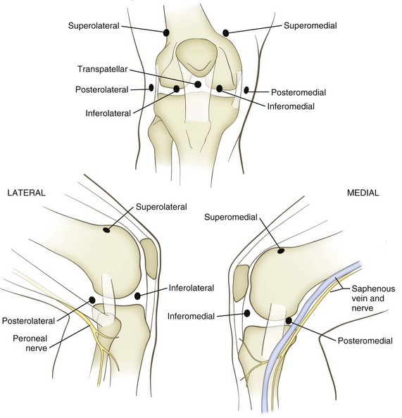
Know Your Knee Anatomy To Code With Confidence Aapc Knowledge Center

Arthroscopic Anatomy Of The Knee Musculoskeletal Key

Cpt code 29883 reports a meniscus repair in.
Arthoscopy medial gutter knee.
Knee Arthroscopy Setup Diagnosis Portals And Approaches Musculoskeletal Key

Figure 4 From Arthroscopic Posteromedial Capsular Release Semantic Scholar

Anatomical Perspective Of The Ankle Arthroscopic Views Download Scientific Diagram

Shotgun Pellet Removal From Medial Meniscus In Left Knee A The Body Download Scientific Diagram
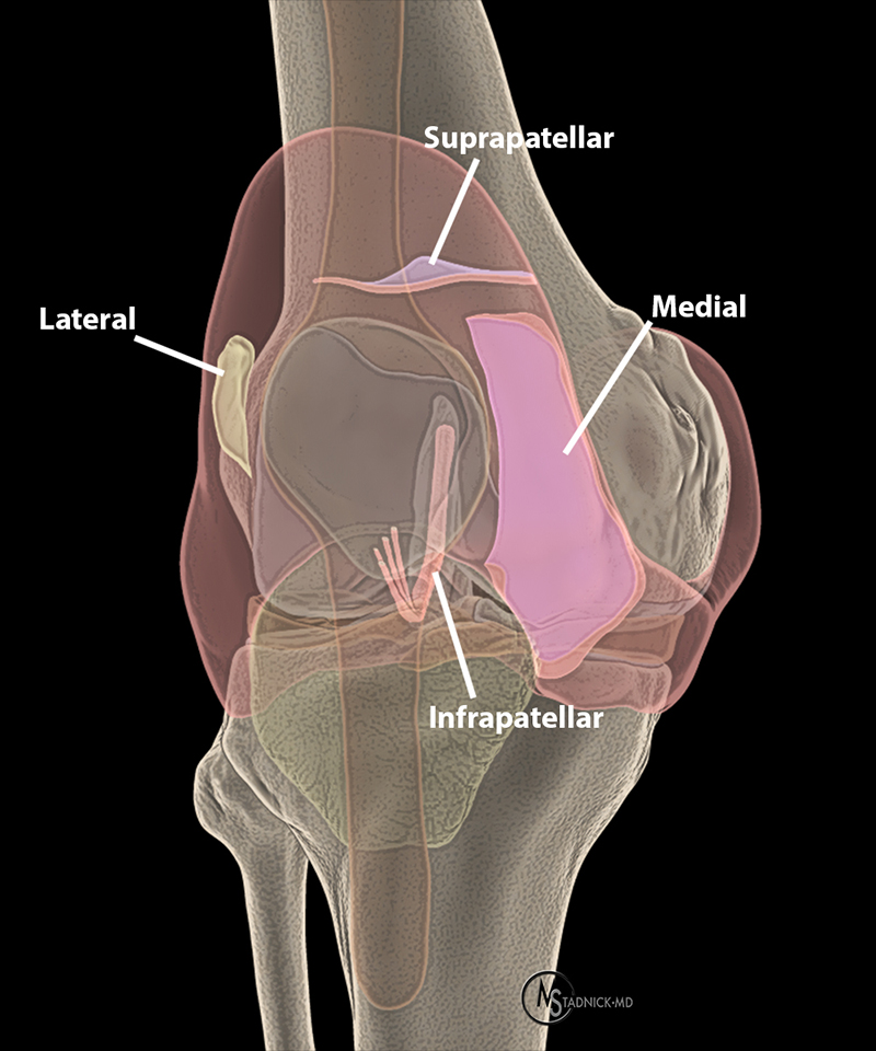
Synovial Plicae Of The Knee Radsource

Aaos Bulletin April 2005
Arthroscopic Versus Open Synovectomy In Rheumatoid Knee
Knee Arthroscopy General Setup Portal Options And How To Manage A Complete Arthroscopic Investigation Springerlink

Case 2 Arthroscopic View Of The Medial Gutter From The Anterolateral Download Scientific Diagram

Mark Clathworthy Patrick Djian Bjorn Engstrom Bent Wulff Jakobsen Ppt Video Online Download

Arthroscopic View Of The Medial Gutter Of A Left Knee Stock Photo Download Image Now Istock

Arthroscopic Medial Menisectomy And Chondroplasy Of Knee Surgical Technique Orthoracle

Arthroscopic Treatment Of Medial Femoral Knee Osteochondral Defect Using Subchondroplasty And Chitosan Based Scaffold Arthroscopy Techniques

Amicus Illustration Of Amicus Surgery Arthroscopic Degenerative Talus Tibial Plafond Synovitis Medial Lateral Gutters Ankle Joint Debrided Shaver Longitudinal Incision Fibular Plate Screws

Pdf Combined Arthroscopic All Inside Repair Of Lateral And Medial Ankle Ligaments Is An Effective Treatment For Rotational Ankle Instability
Sports Medicine Musculoskeletal Key

Case Study Management Of Medial Meniscal Tear And Patellar Osteochondral Damage In A 60 Year Old Female

Knee Arthroscopy With Partial Meniscectomy 29881 Eorif
Https Encrypted Tbn0 Gstatic Com Images Q Tbn 3aand9gcq40mjpiinu01yxzk22jpk0dadzevvvgs2zvl2qnnvpybuvalvv Usqp Cau

Is It Okay If My Knee Makes Noise While Sitting And Standing Quora

Case Study Management Of Ramp Lesion Of The Medial Meniscus With Acl Deficient Left Knee In A 30 Year Old Female
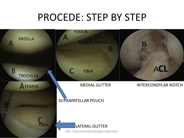
Basics Of Knee Arthroscopy By Dr D P Swami

Knee Arthroscopy Musculoskeletal Key

Left Knee Arthroscopic View Through Anterolateral Portal Showing Download Scientific Diagram
Source : pinterest.com
